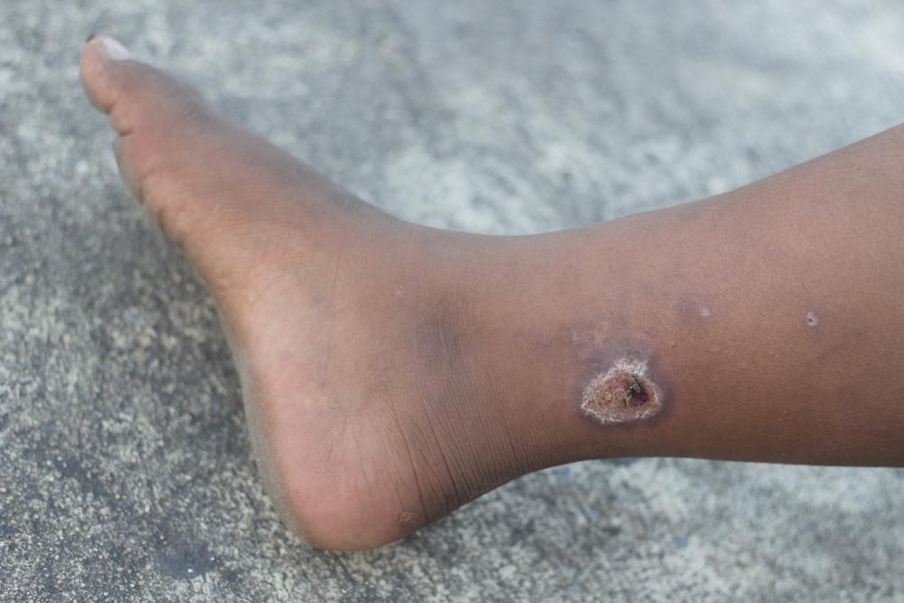
When a patient presents to their podiatrist or wound care specialist with a lower extremity ulcer, the exact type of ulcer might not be readily apparent. Being able to accurately diagnose and identify an ulcer type is important as it helps in the formulation of management. The incidence of diabetic ulcers is steadily increasing due to rising rates of diabetes worldwide. In contrast, venous ulcers also affect a significant population with over 80% of all lower leg ulcers contributed by venous factors.1 On a perfunctory glance, diabetic and venous ulcers appear similar. However, a careful physical examination helps podiatrists differentiate between the two and provide appropriate wound care accordingly.
Location
Venous ulcers are located in the lower extremities, usually below the knee. They are most commonly located near medial malleolus in the "gaiter region". These ulcers are found in the lower limb affected by vascular insufficiency.2
Diabetic ulcers on the other hand are located at pressure points of the foot such as heels, toes, and bony prominences. They are most likely to occur on the areas of thickened callus as punched-out ulcers. However, neurotrophic ulcers caused due to trauma can occur in other locations on the foot as well.
Appearance
Venous ulcers are large, shallow, and have a reddish base. These might present with an excessive amount of wound exudate due to the underlying venous incompetence. The amount of exudate is influenced by the degree of venous valve competency and the presence of other underlying medical conditions like congestive heart failure. Even though wound exudate is a critical component of wound healing, the excess can delay the process. The margins are irregular, and the surrounding skin is discolored due to hemosiderin deposition. The hyperpigmentation around venous ulcers is known as “lipodermatosclerosis”. The skin surrounding the ulcer might also exhibit crusting and eczematous changes. They are associated with swelling and occur in patients with varicose veins.3
Diabetic ulcers are punched-out in appearance. The ulcer bed is variable and might appear red, black, or pink. The ulcer can be of variable thickness but is usually deep involving the skin, tendons, fascia, and even bones. In addition, due to sustained pressure, the skin surrounding diabetic ulcers is thickened and hardened.
Medical History
A thoroughly obtained medical history helps wound care specialists arrive at an accurate diagnosis. The following findings in a patient’s history are strongly suggestive of venous ulceration:
- Presence of varicose veins
- Family history of venous diseases
- Phlebitis
- Prolonged bed rest
- History of recent surgery
- Pregnancy
- Signs and symptoms suggestive of venous insufficiency. These include cramping pains in the legs, itchiness, eczematous changes around the ulcer skin, and swelling.
Some features suggestive of diabetic ulcers include:
- History of diabetes mellitus
- Family history of diabetes
- Neuropathy
- Foot deformity
- Vascular disease
- Previous history of foot ulcers
- Unnoticed foot trauma
A comprehensive medical history can effectively rule out alternative diagnoses and help clinicians optimize wound care depending on the etiology.
Signs and Symptoms
Venous ulcers present with mild pain and aching discomfort. The symptoms can be alleviated by leg elevation and gentle massage of the skin. They also have other symptoms of varicose veins which include swelling, heaviness, and swollen visible veins in the lower limbs. There is also itching in the skin surrounding the venous ulcer.
Diabetic ulcers, in contrast, are painless and present in patients with a history of diabetes mellitus. Neurotrophic ulcers occur due to neuropathic changes caused by hyperglycemia in diabetic patients. Some of the symptoms might include numbness, paraesthesia, and loss of sensation in the lower limbs or feet. Diabetes slows down wound healing and predisposes to infection. Therefore, diabetic ulcers might present with a purulent discharge indicating presence of an infection.4
How to Prevent Venous and Diabetic Ulcers?
Venous and diabetic ulcers both represent a significant burden to the patients and the healthcare system. The incidence of venous and diabetic ulcers can be reduced by following certain preventive strategies. As patients with varicose veins are at an increased risk of venous ulcers, they should wear a compression stocking. Compression stockings are beneficial in reducing the risk of new ulcer formation in patients with an existing venous ulcer.
Diabetic ulcer complications are a leading cause of lower leg amputations. Therefore, to reduce the incidence, patients with diabetes should be informed about the importance of regular foot screening and self-inspection. They should also try to keep their blood sugar levels under control and should wear special protective footwear.
References:
- Collins L, Seraj S. Diagnosis and treatment of venous ulcers. Am Fam Physician. 2010;81(8):989–96.
- Valencia IC, Falabella A, Kirsner RS, Eaglstein WH. Chronic venous insufficiency and venous leg ulceration. Journal of the American Academy of Dermatology. 2001 Mar 1;44(3):401-24.
- Raju S, Neglén P. Chronic venous insufficiency and varicose veins. New England Journal of Medicine. 2009 May 28;360(22):2319-27.
- Frykberg RG. Diabetic foot ulcers: pathogenesis and management. American family physician. 2002 Nov 1;66(9):1655.


.webp)

.avif)