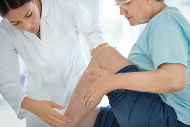
Arterial ulcers are skin lesions formed due to arterial insufficiency or peripheral arterial disease (PAD). They usually develop in the lower limbs, especially the feet and toes. Venous ulcers, on the other hand, are wounds precipitated by blood returning to the veins, primarily due to chronic venous insufficiency. These are ulcers that are more frequent on the calf and ankle.
Venous ulcers are the most common leg ulcers prevalent in elderly people, those with a history of deep vein thrombosis, and those with varicose veins. Arterial ulcers are fewer in number than venous ulcers and are more likely to occur in a person who has factors such as diabetes or cardiovascular diseases. Both of these considerably alter the functions of the patients and their lives, especially the elderly or chronically ill patients.
This problem makes it necessary to distinguish between arterial and venous ulcers. If the wound is misidentified, then the kind of treatment that is administered will not have a positive impact, and healing will be slower, as the patient might get an infection or even lose a limb. Arterial ulcers require treatments that enhance the blood flow in the region, while venous ulcers require treatments that enhance the return of blood to the veins; thus, there is a need to differentiate the two.
Overview of Arterial Ulcers
Causes
Arterial ulcers are formed by ischemia when oxygen and blood cannot reach the tissues. This occurs when the blood vessels become constricted or even occluded, and this is more prevalent in atherosclerosis, where fatty formations develop within the vessels. Peripheral arterial disease (PAD), a disease of the arteries of the limbs with a preference for the lower limbs, is the most common secondary cause of arterial ulcers. Consequently, the tissues are not well supplied with oxygen and nutrients and, as such, do not survive hence the formation of ulcers. In managing arterial ulcers, the essential aspect is to understand that poor circulation is the cause of these ulcers and develop strategies for supplying blood to the legs or arms.
Symptoms
The clinical presentation of arterial ulcers is distinct and often recognizable based on their location and appearance:
Location: Arterial ulcers appear in the regions of the body most distant from the heart where arterial flow is the least. These are seen on the feet, toes, and heels, as well as other areas that experience pressure from shoes, such as the tips of toes or the bony parts of the feet.
Appearance: Arterial ulcers are usually non-tender, non-healing, painless, dry, deep, and have a punched-out appearance with clean margins. The base of the ulcer may appear necrotic, that is, dark, discolored, or black, because the cells are dead, being starved of oxygen.
Pain: Arterial ulcers are typically very tender with or without leg elevation, which occasionally results in additional blood circulation reduction during the night.
Risk Factors
Several factors increase the likelihood of developing arterial ulcers, most of which are linked to atherosclerosis and arterial disease:
Smoking: This is one of the primary reasons why smoking is considered to be a significant contributor to the formation of arterial ulcers since it causes the blood vessels to deteriorate.
Diabetes: Diabetes harms the blood vessels, affecting blood circulation by raising the chances of contraction or blockage in arteries.
Hypertension: Mechanical stress, which contributes to the progression of atherosclerosis, is a significant type of stress on the arterial walls.
High cholesterol: High cholesterol leads to the formation of fatty plaques on the walls of the arteries, narrowing the passage for blood to flow.
Diagnosis
Accurate diagnosis is crucial in managing arterial vs venous ulcers, as the treatments differ greatly depending on the underlying cause. Several diagnostic tools are used to assess blood flow in patients with suspected arterial ulcers:
Ankle-Brachial Index (ABI): This painless procedure measures blood pressure in different body parts, comparing the blood pressure ranges in the ankle with those of the arm. An ABI of less than 0.9 indicates a lack of blood supply to the lower limbs, meaning arterial insufficiency.
Doppler Ultrasound: A Doppler ultrasound evaluates blood circulation in the arteries using sound waves. It helps identify areas such as blockages or narrowing in the blood vessels.
Angiography: Angiography is an imaging test similar to angioplasty that enables doctors to precisely locate blockages and areas of atherosclerosis, often using contrast dye.
Treatment
The treatment of arterial ulcers focuses on improving blood flow to the affected area, managing symptoms, and promoting wound healing:
Revascularization Procedures: Most importantly, surgical interventions, such as revascularization, may be required to restore blood flow in the affected area. This may be achieved through angioplasty – where the arteries are opened with the help of a balloon or bypass surgery – where the blood flow is directed around the blockage.
Wound Care: Good ulcer dressing is vital in managing arterial ulcers. Surgery may be necessary to carry out a debridement process to remove dead or infected tissue; specific meticulous dressings are applied to protect the ulcer, support the moist healing environment, and prevent infections.
Pain Management and Lifestyle Changes: Pain management is essential since arterial ulcers are excruciating and require proper patient comfort management. That is why managing these risk factors is crucial to abstaining from the growth of further ulcers. It comprises quitting smoking, controlling diabetes and hypertension, and reducing cholesterol levels, to name but a few.
Overview of Venous Ulcers
Causes
Venous ulcers are a result of slow circulation of blood in the veins, which is known as venous insufficiency. This is due to inadequate blood returning into the heart through the veins, which results in high pressure in the veins. It leads to dilation of the veins and damage to the valves, which allow fluid and protein to leak into tissues, causing skin breakdown and ulcers. The prevalence of risk factors for venous ulcers is best known with chronic venous insufficiency, also called CVI, a long-term condition where the veins in the legs cannot efficiently return blood to the heart.
Symptoms
Venous ulcers present a distinct set of symptoms, which can help differentiate them from arterial ulcers:
Location: Venous ulcers are commonly found in the lower legs, specifically in the ankle area, also known as the ‘gaiter area.’
Appearance: Venous ulcers usually have partial thickness, and the ulcer margins are often poorly defined, irregular, and jagged. The wound bed is commonly described as ‘weeping’ or ‘oozing’ and has features of excess exudate. Arterial ulcers are usually dry, but venous ulcers have a congestive background, and therefore the fluid leaks through the damaged veins.
Pain: The pain is often characterized as an ache or a throbbing pain. It worsens when the patient is on their feet or walking because these activities raise pressure in the legs. Leg pain is soothed by raising the legs to decrease the blood flow, lowering pressure on the veins.
Risk Factors
Several factors increase the risk of developing venous ulcers, most of which are related to conditions that impair venous circulation:
Varicose Veins: Varicose veins are swollen and twisted, which can cause slow blood circulation and the development of a possible chronic ulcer.
Deep Vein Thrombosis (DVT): Experience of DVT, which is the formation of blood clots in the deep veins of the lower limbs, predisposes people to chronic venous insufficiency that results in the formation of venous ulcers.
Obesity: Obesity increases the pressure on the veins, making it difficult for blood to flow back to the heart, causing venous insufficiency.
Diagnosis
Proper diagnosis of venous ulcers is essential to differentiate them from arterial ulcers and to ensure appropriate treatment. Diagnostic tests focus on evaluating venous function and blood flow:
Duplex Ultrasound: Duplex ultrasound is the most common test for evaluating blood flow in the veins. Its ability to give the physician a picture of the veins and the speed at which blood flows through them further enhances this evaluation.
Venography: In venography, a contrast dye is introduced into the veins, and then X-ray images are taken to determine whether there are any blockages or abnormalities.
Treatment
The treatment of venous ulcers focuses on improving venous circulation, reducing pressure in the veins, and promoting wound healing:
Compression Therapy: Compression therapy is often used as a treatment. It applies pressure on the leg, thus increasing blood circulation and preventing the accumulation in the lower part of the body. It also plays a role in treating ulcers due to increased oxygen delivery to the tissues.
Elevation and Wound Care: Patients should raise their legs above the heart level several times daily, reducing pressure on the veins and promoting blood circulation. Cleaning and dressing wounds to prevent infection and encourage faster healing is vital in managing venous ulcers. Specific dressings and wound management practices that control exudate and moisten the wound are also necessary.
Surgical Intervention: If conservative interventions do not yield positive results, chances are high that one will need surgery. Operations such as ligation and stripping, where the varicose veins are stripped, or sclerotherapy, where a solution is injected into the affected veins to seal them, aid in the correct venous flow to avoid the recurrence of ulcers.
Key Differences Between Arterial and Venous Ulcers
Understanding the critical differences between arteria ulcers vs venous ulcers is essential for accurate diagnosis and treatment, as these two types have distinct causes, symptoms, and treatment approaches. Here’s a comparison of the primary distinctions:
Etiology
Arterial Ulcers:
Arterial ulcers are caused by arterial insufficiency, which occurs when the arteries cannot deliver sufficient oxygen-rich blood to the tissues. This is often due to atherosclerosis, where plaque builds inside the arteries, leading to blockages and reduced blood flow. Arterial ulcers are typically seen in patients with peripheral arterial disease (PAD), where blood flow to the lower limbs is compromised.
Venous Ulcers:
Venous ulcers, conversely, are caused by venous insufficiency, a condition in which the veins cannot effectively return blood from the lower limbs to the heart. Chronic venous insufficiency (CVI) leads to increased pressure in the veins, which damages the vein walls and valves. Over time, this venous pressure causes fluid to leak into surrounding tissues, resulting in skin breakdown and ulcer formation.
Location
Arterial Ulcers:
Arterial ulcers are typically found on the feet, toes, and pressure points, where blood flow is already limited, and minor trauma can exacerbate the lack of oxygen supply. These ulcers are commonly located on the tips of the toes, the heels, and over bony prominences, such as the ankles. Their location is usually distal (far from the heart), in areas where blood supply is most diminished.
Venous Ulcers:
Venous ulcers primarily develop in the lower legs, particularly near the ankle in the "gaiter area," the region just above the ankle and below the calf. Due to gravity, blood pooling tends to occur in this location, especially in patients with venous insufficiency. These ulcers rarely occur on the feet or toes, distinguishing them from arterial ulcers.
Pain Characteristics
Arterial Ulcers:
The pain associated with arterial ulcers is often described as severe and intense due to the lack of oxygen reaching the tissues. This pain typically worsens when the leg is elevated as blood flow to the affected area becomes even more restricted. Patients with arterial ulcers often find relief by lowering the affected limb to improve circulation.
Venous Ulcers:
Venous ulcers usually cause mild to moderate pain, often described as aching or throbbing. The pain worsens after standing for long periods, as blood pools in the lower extremities, increasing pressure. Unlike arterial ulcers, the pain from venous ulcers is relieved by elevating the legs, which helps reduce venous pressure and improves blood flow.
Appearance
Arterial Ulcers:
Arterial ulcers have a characteristic appearance that sets them apart from venous ulcers. They are typically dry, deep, and have well-defined edges. The base of the ulcer is often necrotic, with dead tissue that appears black or brown due to the lack of oxygen and blood supply. These ulcers often have a "punched-out" appearance, with a clear separation between the ulcer and the surrounding skin.
Venous Ulcers:
In contrast, venous ulcers tend to be moist and shallow, with irregular edges. They are often covered in a layer of yellow or brown fibrin, a protein involved in the clotting process. Venous ulcers are typically exudative, producing a significant amount of fluid (exudate), making them appear wet and oozing. The surrounding skin may also appear macerated due to the constant exposure to moisture.
Skin Surrounding Ulcers
Arterial Ulcers:
The skin around arterial ulcers often exhibits signs of poor circulation. The surrounding skin may appear cool, shiny, and atrophic (thin and fragile). Hair loss is expected in the affected area, and the skin may look pale or bluish due to inadequate blood flow. Over time, the tissue may become ischemic, leading to more significant damage and a higher risk of infection.
Venous Ulcers:
The skin around venous ulcers typically shows signs of chronic venous disease, such as hyperpigmentation (darkening of the skin due to the leakage of blood pigments). Other standard features include stasis dermatitis (inflammation of the skin due to venous insufficiency) and edema (swelling). The skin may also become thickened, leathery, and firm. In advanced cases, lipodermatosclerosis, a condition where the skin becomes hardened and scarred, may develop.
Treatment Strategies for Arterial and Venous Ulcers
The treatment strategies for arterial vs venous ulcers differ significantly due to their underlying causes. While arterial ulcers result from poor blood flow through the arteries, venous ulcers are caused by inadequate blood return through the veins. Therefore, the primary goal in treating arterial ulcers is to restore proper circulation, whereas the focus for venous ulcers is improving venous return and managing swelling. Below are the specific approaches for treating each type of ulcer.
Improving Arterial Blood Flow
The cornerstone of treating arterial ulcers is restoring blood flow to the affected area. This is critical to promote healing, as arterial ulcers are caused by ischemia (reduced blood supply). Various revascularization procedures are used to improve circulation:
Angioplasty: A minimally invasive procedure where a balloon widens the narrowed or blocked arteries, restoring blood flow to the affected limb. A stent may also be placed to keep the artery open.
Bypass Surgery: In more severe cases of arterial insufficiency, bypass surgery is performed to reroute blood flow around a blocked artery. A graft creates a new pathway for blood to reach the tissues.
Pain Control and Wound Care
Pain management is crucial for patients with arterial ulcers, as these ulcers are often very painful due to the lack of oxygen reaching the tissues. Pain is typically worse when the limb is elevated. Pain relief strategies include:
Medications: Analgesics, including non-steroidal anti-inflammatory drugs (NSAIDs) and opioids, may be prescribed for pain relief. Topical anesthetics can also be used for localized pain control during wound care.
Wound care involves:
Debridement: Removing dead or necrotic tissue from the ulcer promotes healing and reduces the risk of infection.
Moisture balance: While arterial ulcers are typically dry, an appropriate moisture balance is essential for wound healing. Specialized dressings like hydrocolloids may protect the wound and keep it moist.
Infection control: Patients with arterial ulcers are at high risk of infection due to poor circulation. If signs of infection are present, antibiotics may be required.
Lifestyle Changes
Managing risk factors and adopting lifestyle changes is critical for preventing further arterial ulcer development and improving overall vascular health:
Smoking cessation: Smoking significantly worsens arterial disease by narrowing blood vessels and reducing circulation. Quitting smoking is essential for improving blood flow.
Control of comorbidities: Managing conditions such as diabetes, hypertension, and high cholesterol is vital to prevent the progression of arterial disease. This can involve dietary changes, exercise, and medications to control blood pressure, blood sugar, and cholesterol levels.
Treatment Strategies for Venous Ulcers
Compression Therapy
Compression therapy is the cornerstone of treatment for venous ulcers. Since these ulcers are caused by poor venous return and fluid buildup in the lower legs, applying pressure helps improve blood flow and reduces swelling. Compression therapy can take several forms:
Compression stockings: These specially designed garments apply graduated pressure to the legs, helping push blood back toward the heart and reduce venous congestion.
Compression wraps or bandages: These are often used in more severe cases to provide consistent pressure on the ulcerated area and manage excessive exudate.
Intermittent pneumatic compression devices: Sometimes, these devices may deliver controlled pressure at intervals to enhance blood flow.
Elevation and Wound Care
Elevation of the legs helps reduce venous pressure and swelling, promoting healing. Patients are advised to keep their legs above the heart level several times a day for at least 30 minutes to encourage proper blood flow and decrease fluid buildup.
Wound care for venous ulcers focuses on the following:
Moist wound healing: Because venous ulcers tend to be moist with exudate, proper dressings are essential to manage the fluid and promote healing. Dressings such as alginates or foam may absorb excess fluid while keeping the wound bed moist.
Debridement: If necrotic tissue is present, debridement may be necessary to clean the wound and stimulate healing. This is especially important if the ulcer becomes infected.
Addressing Venous Insufficiency
Treating the underlying venous insufficiency is crucial to prevent the recurrence of venous ulcers. Several procedures may be used to correct dysfunctional veins:
Sclerotherapy: In this procedure, a solution is injected into the affected veins, causing them to collapse and close. This helps redirect blood flow to healthier veins, reducing venous congestion.
Endovenous Ablation: This minimally invasive technique uses heat from lasers or radiofrequency energy to close off the damaged veins, improving venous circulation.
Vein Stripping or Ligation: In more severe cases, surgery may be required to remove or tie off the affected veins, preventing further blood pooling.
Conclusion
Differentiating between arterial vs venous ulcers is crucial for appropriate treatment, as each type of ulcer requires a distinct therapeutic approach. Arterial ulcers, caused by poor blood flow due to arterial insufficiency, demand strategies focused on restoring circulation, such as revascularization, pain control, and lifestyle changes. Venous ulcers, on the other hand, result from venous insufficiency and are managed primarily through compression therapy, elevation, and procedures to improve venous return. Accurately identifying the type of ulcer ensures that patients receive targeted treatment, preventing complications and promoting faster healing.
Early intervention and preventive strategies are essential to improving patient outcomes in arterial and venous ulcers. Prompt diagnosis, risk factor management, and lifestyle modifications can significantly reduce the likelihood of ulcer formation or recurrence. A multidisciplinary approach involving vascular specialists, wound care experts, and primary care physicians is often necessary for long-term management. This collaborative care model addresses the underlying vascular conditions while providing comprehensive wound care, ensuring better outcomes and improved quality of life for patients.

.webp)
.avif)